Home
/ Human Neck Anatomy Diagram / Human Neck Anatomy High Resolution Stock Photography And Images Alamy - In their first year, residents should be well versed with normal radiographs, ultrasound and ct anatomy followed.
Human Neck Anatomy Diagram / Human Neck Anatomy High Resolution Stock Photography And Images Alamy - In their first year, residents should be well versed with normal radiographs, ultrasound and ct anatomy followed.
Human Neck Anatomy Diagram / Human Neck Anatomy High Resolution Stock Photography And Images Alamy - In their first year, residents should be well versed with normal radiographs, ultrasound and ct anatomy followed.. Anatomynote.com found common carotid artery diagram from plenty of anatomical pictures on the internet. They are attached to the femur (thighbone), tibia (shinbone), and fibula (calf bone) by fibrous tissues called ligaments. Veins and arteries of the neck 9 photos of the veins and arteries of the neck activate javascript arteries in the neck diagram, common carotid artery branches, external carotid artery function, how many carotid arteries, left common carotid artery function, the left common carotid artery supplies blood to the. In their first year, residents should be well versed with normal radiographs, ultrasound and ct anatomy followed. See anatomy of the head and neck stock video clips.
The neck is the start of the spinal column and spinal cord. The anatomy of the head and neck of the human body, including the bones, muscles, blood vessels, nerves, glands, nose, mouth, and throat. 604), three to six in number, are placed beneath the body of the mandible in the submaxillary triangle, and rest on the superficial surface of the submaxillary salivary gland.one gland, the middle gland of stahr, which lies on the external maxillary artery as it turns over the mandible, is the most constant of the series; In this article, you will read about each specific area of the throat as well as its surrounding structures. Broadly considered, human muscle—like the muscles of all vertebrates—is often divided into striated muscle, smooth muscle, and cardiac muscle.
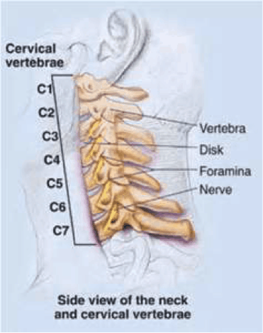
Neck Anatomy Pictures Bones Muscles Nerves from www.healthpages.org The content of the neck is grouped into 4 neck spaces, called the compartments. The anatomy of the head and neck of the human body, including the bones, muscles, blood vessels, nerves, glands, nose, mouth, and throat. Contains cervical vertebrae and postural muscles. The neck is the start of the spinal column and spinal cord. 604), three to six in number, are placed beneath the body of the mandible in the submaxillary triangle, and rest on the superficial surface of the submaxillary salivary gland.one gland, the middle gland of stahr, which lies on the external maxillary artery as it turns over the mandible, is the most constant of the series; The axial skeleton and the appendicular skeleton. The head rests on the top part of the vertebral column, with the skull joining at c1. These bones are arranged into two major divisions:
The neck contains seven of.
The throat is positioned in the anterior part of the neck. Anterior, lateral and posterior groups, based on their position in the neck.the musculature of the neck is further divided into more specific groups based. Diagram of neck bones, human neck bones diagram, neck bones diagram, bone, diagram of neck bones, human neck bones diagram, neck bones diagram. In this article, you will read about each specific area of the throat as well as its surrounding structures. Muscle head anatomy vocal organ diagram female neck anatomy neck wireframe head neck human anatomy head artery anatomy face pharynx vector neck degree head anatomy 3d. For more anatomy content please follow us and visit our website: The nasopharynx is the uppermost part of the pharynx and extends down from the base of the skull to the nasal passages, hence the name. There are many muscles around the neck that help to support the cervical spine and allow you to move your head in different directions. Some important structures contained in or passing through the neck include the seven cervical vertebrae and enclosed spinal cord the jugular veins and carotid arteries part of the. Human muscle system, the muscles of the human body that work the skeletal system, that are under voluntary control, and that are concerned with movement, posture, and balance. Human body anatomy background of a male and female muscle anatomy of the face neck and shoulder medical image reference of human anatomy stock photo alamy / 6 best images of printable worksheets muscle anatomy blank head and neck muscles diagram muscular system diagram worksheet and label muscles worksheet. Contains glands ( thyroid, parathyroid, and thymus ), the larynx, pharynx and trachea. We think this is the most useful anatomy picture that you need.
The nasopharynx is the uppermost part of the pharynx and extends down from the base of the skull to the nasal passages, hence the name. Neck human body anatomy head and neck anatomy reference body diagram muscles of the neck neck muscle anatomy muscle structure anatomy organs. Anterior, lateral and posterior groups, based on their position in the neck.the musculature of the neck is further divided into more specific groups based. The external jugular vein (v. The anatomy of the head and neck of the human body, including the bones, muscles, blood vessels, nerves, glands, nose, mouth, and throat.
Human Body Diagrams Wikimedia Commons from upload.wikimedia.org .and neck muscles diagram can help you study and research.arteries in the neck diagram common carotid artery branches external carotid artery function how many. Posted in anatomy, body parts | tagged anatomy of neck, human neck, neck anatomy, neck chart, neck diagram, neck diagram with labels, neck explained, where is neck. Labeled anatomy chart of neck and shoulder muscles on black background labeled human anatomy diagram of man's neck and shoulder muscles in an anterior view on a black background. For more anatomy content please follow us and visit our website: Contains cervical vertebrae and postural muscles. The throat is a complex part of the body with many structures that surround it that are very important. There are many muscles around the neck that help to support the cervical spine and allow you to move your head in different directions. The external jugular vein (v.
Related posts of anatomy of neck muscles diagram anatomy of human pancreas.
Anterior, lateral and posterior groups, based on their position in the neck.the musculature of the neck is further divided into more specific groups based. The nasopharynx is the uppermost part of the pharynx and extends down from the base of the skull to the nasal passages, hence the name. Posted on june 7, 2016 by admin. Human muscle system, the muscles of the human body that work the skeletal system, that are under voluntary control, and that are concerned with movement, posture, and balance. They are attached to the femur (thighbone), tibia (shinbone), and fibula (calf bone) by fibrous tissues called ligaments. Über 7 millionen englischsprachige bücher. The throat is a complex part of the body with many structures that surround it that are very important. The thyroid gland is located in the neck below the thyroid cartilage, or adam's apple. Some important structures contained in or passing through the neck include the seven cervical vertebrae and enclosed spinal cord, the jugular veins and carotid arteries, part of the esophagus, the larynx The external jugular vein (v. Broadly considered, human muscle—like the muscles of all vertebrates—is often divided into striated muscle, smooth muscle, and cardiac muscle. Neck vessel anatomy (page 1). Related posts of diagram of the neck anatomy veins and arteries of the neck.
Raj md on august 13th, 2018. The axial skeleton and the appendicular skeleton. We hope this picture common carotid artery diagram can help you study and research. The neck contains seven of. Anatomynote.com found common carotid artery diagram from plenty of anatomical pictures on the internet.
Head And Neck Anatomy Human Body Organ V Jugularis Externa Head Anatomy Abdomen Png Klipartz from c0.klipartz.com Broadly considered, human muscle—like the muscles of all vertebrates—is often divided into striated muscle, smooth muscle, and cardiac muscle. Human muscle system, the muscles of the human body that work the skeletal system, that are under voluntary control, and that are concerned with movement, posture, and balance. Contains cervical vertebrae and postural muscles. The head rests on the top part of the vertebral column, with the skull joining at c1. The external jugular vein (v. This atlas is part of a growing encyclopedic resource covering human anatomy and includes image libraries, presentations, videos, interactive self assessment, and brand new 3d content from ehuman digital anatomy. The neck contains seven of. Anatomy human neck muscle diagram & pictures.
Anatomy human neck muscle diagram & pictures.
Anatomy of back of human neck anatomy of the back and neck anatomy of the back of the neck anatomy of the back of the neck muscles anatomy of the back of your. .and neck muscles diagram can help you study and research.arteries in the neck diagram common carotid artery branches external carotid artery function how many. The neck muscles, including the sternocleidomastoid and the trapezius, are responsible for the gross motor movement in the muscular system of the head and neck. See anatomy of the head and neck stock video clips. Contains glands ( thyroid, parathyroid, and thymus ), the larynx, pharynx and trachea. Working in pairs on the left and right sides of the body, these muscles. Related posts of anatomy of neck muscles diagram anatomy of human pancreas. For more anatomy content please follow us and visit our website: Human muscle system, the muscles of the human body that work the skeletal system, that are under voluntary control, and that are concerned with movement, posture, and balance. Muscles of the neck (musculi cervicales) the muscles of the neck are muscles that cover the area of the neck hese muscles are mainly responsible for the movement of the head in all directions they consist of 3 main groups of muscles: Human body anatomy background of a male and female muscle anatomy of the face neck and shoulder medical image reference of human anatomy stock photo alamy / 6 best images of printable worksheets muscle anatomy blank head and neck muscles diagram muscular system diagram worksheet and label muscles worksheet. Labeled anatomy chart of neck and shoulder muscles on black background labeled human anatomy diagram of man's neck and shoulder muscles in an anterior view on a black background. Contains cervical vertebrae and postural muscles.
The content of the neck is grouped into 4 neck spaces, called the compartments neck anatomy diagram. Muscles of the neck (musculi cervicales) the muscles of the neck are muscles that cover the area of the neck hese muscles are mainly responsible for the movement of the head in all directions they consist of 3 main groups of muscles:

